Special Stain - Martius Scarlet Blue
- Fibrin Staining
- Martius Scarlet Blue Technique
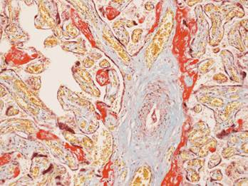
- Placenta MSB staining
The MSB method falls into the category of trichrome stains, by implication three dyes are employed. The general rule in trichrome staining is that the less porous tissues are coloured by the smallest dye molecules, and whenever a dye of larger molecular size is able to penetrate it, it will always do so at the expense of the smaller molecules.
MSB is a trichrome method for selective demonstration of fibrin. Fibrin is stained using crystal scarlet solution, while methyl blue is used to stain the collagen. Red blood cells are stained yellow by the picric acid.
The Method
- Use appropriate control section.
- Take sections to water
- Stain in Celestin blue (2 mins), Take to water then Haematoxylin (2mins) sequence
- Differentiate in 1% acid alcohol if necessary.
- Wash in tap water. Blue in Lithium Carbonate
- Rinse in 95% alcohol.
- Stain in Martius Yellow solution - 5 minutes.
- Rinse in distilled water.
- Stain in Brilliant Crystal Scarlet 6R solution - 10 minutes.
- Rinse in distilled water.
- Treat with Phosphotungstic Acid - 2 minutes. Check microscopically. Repeat, if necessary.
- Rinse in distilled water.
- Counterstain in Methyl blue solution diluted 1:10 with 1% acetic acid - 2 minutes. Check microscopically. If pale, repeat.
- Rinse in 1% acetic acid.
- Dehydrate, clear and mount
Result
Fibrin |
Red |
Collagen |
Blue |
Muscle |
Red |
RBCs |
Yellow |
Below is an image of a successful MSB that was used on a section of placenta looking for fibrin.
Photo x10 MSB