Special Stains - Toludine Blue
- Metachromasia - Mast Cells
- Toludine Blue Technique
Mast cells are found in the connective tissue and their cytoplasm contains granules composed of heparin and histamine. Toluidine blue stains mast cells red-purple and the background blue . tissue elements staining a different colour from the dye solution , is due to the pH, dye concentration and temperature of the basic dye. Blue or violet dyes will show a red colour shift, and red dyes will show a yellow colour shift with metachromatic tissue elements.
1. Take sections to water.
2. Flood section with 0.1% Tol.blue in 0.1% borax for 5-10 Seconds
3. Wash in tap water.
4. Differentiate by washing in spirit and water until pale background and purple mast cells are visible
5.Wash, dehydrate, clear and mount
Results
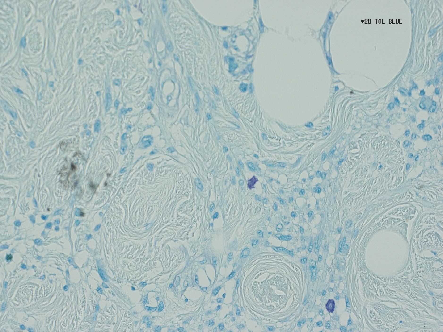
Mast cells - red-purple
Background - pale blue
The results can be seen below, the mast cells are shown up clearly in reddish purple while the background is pale to provide better contrast. This stain is one of the best examples of a metachromatic stain, where the same colour of dye has a differenting effect on the different types of tissue
Photo – Toludine Blue x10