
Special Stains - Gram
- Bacteria - Gram Positive or Negative
- Gram staining Technique
- Gram staining History
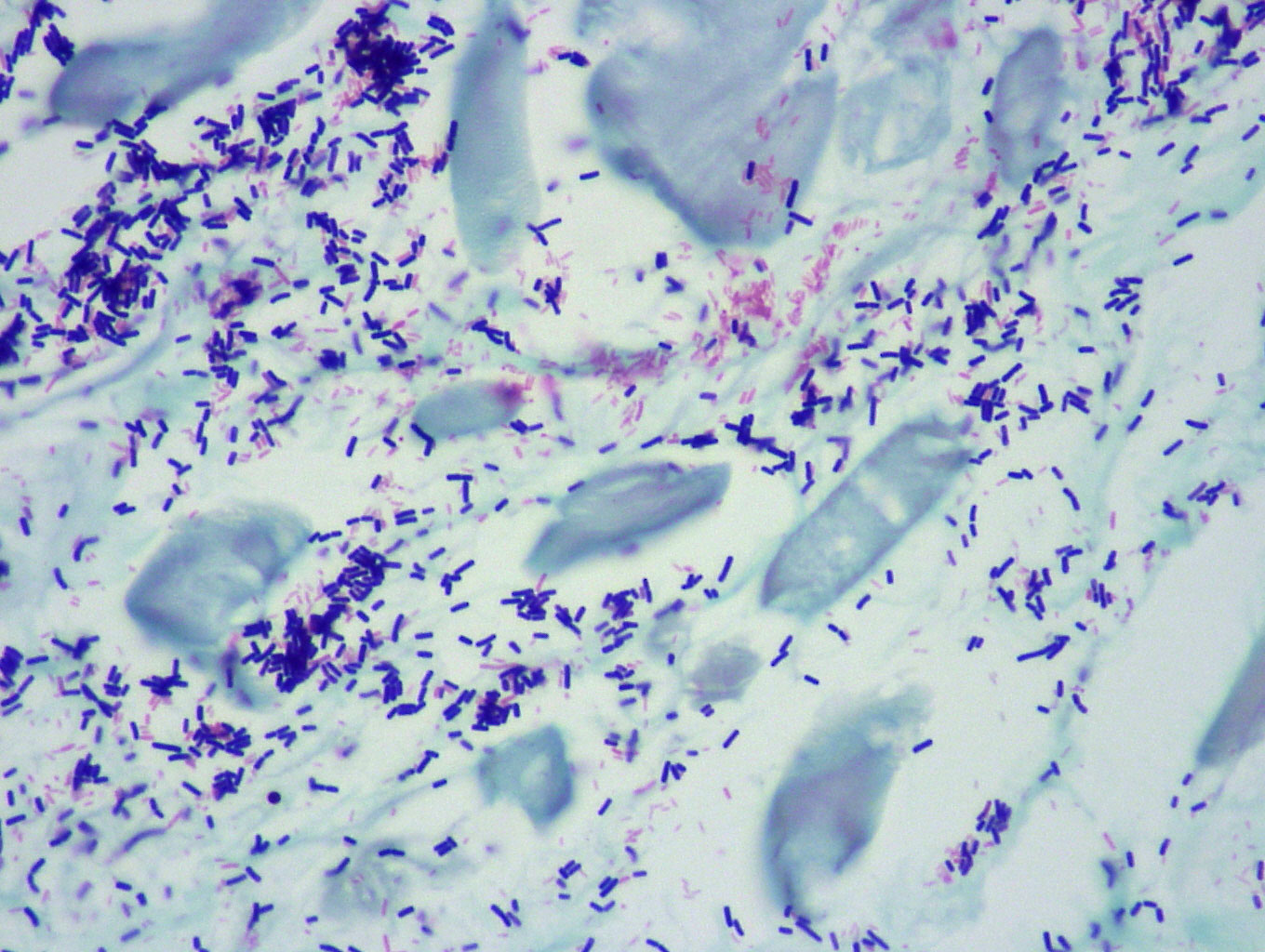
This is the main stain used for staining bacteria. It is a very useful stain as it stains up most bacteria and allows differentiation of gram positive and gram negative bacteria. Bacteria can be classified as either Gram-positive or Gram-negative, based on the composition of the cell wall and its permeability to dyes or stains. The gram stain does this by first overstraining them with crystal violet and then washing out the stain until only the gram positive bacteria retain it as they resist decolourisation by acetone and so are stained blue. Gram negative bacteria are then counterstained with neutral red. This technique is now widely used in histology but also in microbiology as well.
Below is the technique for performing the gram stain.
- Take sections to water
- Stain in ammonium oxalate crystal violet solution for 3 minutes
- Wash in water
- Treat with Grams iodine for 3 minutes
- Wash in water
- Decolourise in acetone until section appears straw coloured.
- Wash in water
- Counterstain in neutral red solution for 5 minutes.
- Dehydrate, clear and mount
Results
Gram positive Organisms |
Blue / Violet |
Gram negative Organisms |
Red |
Nuclei |
Red |
Photo - x10 Gram Stain
The gram staining technique derived its name from the scientist Hans Christian Gram, who developed the technique alongside Carl Friedländer in Berlin hospital, while he worked in the morgue. The technique was created to enable bacteria to be seen more readily in stained sections of lung tissue.He published his method in 1884. Later on it became almost the gold standard technique of of distinguishing one type of bacterium from another