Histological Techniques - Haematoxylin & Eosin
- What is haematoxylin and eosin?
- H&E Staining
- Regressive or Progressive
- Haematoxylin types
The Haematoxylin and eosin staining involves two main dye chemistry processes. The first process is carried out by dying the tissue in Harris haematoxylin, which then will overstain the tissue to allow complete penetration by the solution. The haematoxylin is a basic dye with a + charge to it, so it will stain up the negatively charged nucleic acids in the nuclei. Then the tissue is taken through water before going into acid alcohol to remove some of the haematoxylin which had overstained the tissue. Afterwards it goes through water again before being blued in lithium carbonate, which alters the haematoxylin dyed tissue colouration from purple to blue.
Finally after all this, the tissue undergoes the second part of the procedure. It is then is counter stained using the other main dye Eosin which colours the non nuclei part of the tissue a pink/red colouration to allow a contrast. This is achieved as Eosin is an acid dye, with a negative charge which will stain up + charged areas of the tissue eg cytoplasm. Then the slide finally goes through to water, then dehydrated in alcohol before being cleared in xylene and mounted in a CV mountant agent, to acheive better resolution under the microscope.
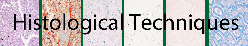
Below are the results of missing out a step in the process, with the skin tissue to demonstrate the importance of the haematoxylin and eosin staining steps. The results are shown below.
Photo 1 – x10 No haematoxylin section Photo 2 – x10 No lithium carbonate
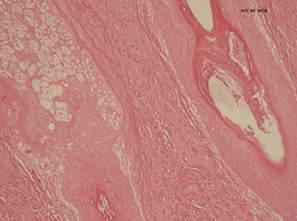
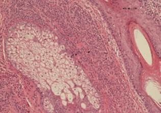
Photo 3 – x10 No acid alcohol Photo 4 –x10 No Eosin
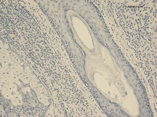
Photo 5 – Normal H&E picture
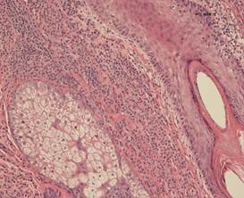
The different stages of the Haematoxylin and eosin staining are all necessary for the production of the expected staining result. In the 1st photo, without the haematoxylin step, the nuclei are unstained, while in the 2nd photo without lithium carbonate stage of bluing, the nuclei are darker purple colouration and the stain is darker in general. In the 3rd photo, lack of acid alcohol step to differentiate the haematoxylin lead to the production of much darker staining of the tissue in general as well. While in the 4th photo without the eosin, only the nuclei are visible and no counter stain means that the rest of the tissue is not visible under these circumstances. The final photo shows what the H&E looks like with all the steps performed.
In H&E staining, there are two main variations used.
Progressive
The first version uses a progressive staining where the tissue is stained with haematoxylin then blued with lithium carbonate without the use of acid alcohol as it is not required as the haematoxylin is applied until the desired staining is achieved, and then it is completed. An example of this is is Mayer haematoxylin.
Regressive
While the second version is regressive staining. This is where the tissue is overstained with haematoxylin, which is then removed using acid alcohol before it is blued using lithium carbonate. In the regressive staining, it is very important to differentiate as without it, the staining will be too dark. An example of this type of staining is Harris haematoxylin staining, which is the most commonly used Haematoxylin. The technique for carrying this out is below.
1. Place section in Xylene to dewax. 5 minutes
2. Wash in 100% Ethanol 30 seconds
3. Wash in IMS 30 seconds
4. Wash in running water 2-5 seconds
5. Place in Haematoxylin 5 minutes
6. Wash in water.
7. Differentiate in 1% Acid Alcohol 2 to 5 seconds.
8. Wash in water.
9. Then Blue inLithium Carbonate.
10. Check nuclear stain microscopically before proceeding, adjust if necessary.
11. Wash in water, rinse in IMS.
12. Stain in Eosin 30 seconds.
13. Wash in IMS then Dehydrate, clear and mount.
Results
Nuclei |
Blue |
Cytoplasm, connective tissues, RBCs |
Pink/Red |
There are many Different variations of haematoxylin, they all however have a similar make up as shown in the table below.
| 3 main constituents of Harris haematoxylin and their function | ||||||||||||||
|---|---|---|---|---|---|---|---|---|---|---|---|---|---|---|
| Constituents | Function | |||||||||||||
| Haematein | Oxidised product of haematoxylin which act as the stain, however it requires a metal mordant due to its – charge which would prevent dying of – charged nucleic acids. | |||||||||||||
| Mercuric Oxide | Oxidising agent used on haematoxylin to produce the haematein | |||||||||||||
| Potassium Alum | Used as the metal mordant in Harris haematoxylin. This gives the haematein a better affinity for the tissue when in use. Other mordants are used in other versions eg Tungsten or Iron. | |||||||||||||