Histological Techniques - What is histology?
- What is Histology?
- Histology History
- Histology Staining
- Histology Table of contents
So what is histology?
The definition of Histology is the study of anatomy of animals and plants down to the cellular level. This involves analysis of biological tissue using microscopy to look at specimens carefully prepared using special processes called "histological techniques".
Histological techniques or Processes
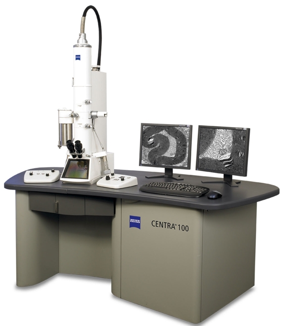
It is performed by first using process called fixation of the tissue using a suitable fixative, mostly 10% neutral buffered formalin. Then the tissue then undergoes processing, mainly to paraffin wax. After this it is Embedding, again mainly into paraffin wax to form a block. Then the tissue block is sectioned using Microtomy. This allows examinination of a thin slice of tissue, which picked up on a slide is stained using normally a Haematoxylin and eosin stain so it can be examined under a light microscope or if you need to examine further into the tissue you can go as far as using an electron microscope which is covered more fully under Electron microscopy. The ability to visualize or differentially identify microscopic structures is frequently enhanced through the use of histological stains also known as special stains or immunohistochemistry or immunofluorescence. Sometimes if a diagnosis is urgent a Frozen section can be performed, often used in hospitals in operations for thyroid or ureter identification.
Histology staff - Who carry out this work?
In laboratories it would normally be Biomedical scientists or healthcare support staff who carry out the histology procedures. They require high levels of training to be competent in all the procedures, only possible for Biomedical scientist staff members after completion of a Registration portfolio to allow registration with the health care professions council (HCPC). Later on further developement to specialise in this area requires completion of a specialist portfolio or diploma with the full name specialist portfolio in cellular pathology. To further specialise or move into managerial roles the staff may complete a Masters in biomedical science or Higher specialist diploma.
The Beginnings of Histology
The journey of histology, a crucial branch of biology, began in 1665 when English microscopist Robert Hooke made a groundbreaking discovery. While examining a thin slice of cork with his rudimentary microscope, Hooke observed the existence of cells, identifying them as the fundamental units of life. However, it's important to note that what Hooke saw were actually the cell walls of dead plant cells. This discovery laid the groundwork for the development of cell theory, a cornerstone of modern biology.
19th Century Breakthroughs
Histology made significant strides in the 19th century, culminating in the 1906 Nobel Prize in Physiology or Medicine being awarded to two notable histologists, Camillo Golgi and Santiago Ramón y Cajal. Their contributions to neuroscience were monumental, albeit rooted in differing interpretations of neural structures in the brain. Golgi was recognized for inventing a revolutionary staining technique, now known as the "Golgi stain," which allowed for detailed visualization of nerve cells and networks. Cajal, on the other hand, won acclaim for his neuron doctrine, which accurately described how neurons are separate entities rather than continuous networks. These interpretations were based on observations made possible by Golgi’s staining method.
Advancements in Staining Techniques
Since the early 20th century, the repertoire of stains used in histological laboratories has expanded significantly. From the most common haematoxylin and eosin stain, used to differentiate between cell and tissue types, to more specialized stains, each has a unique role. For instance, the "alcian blue" stain is utilized for mucin detection, crucial in identifying certain types of cancer cells. Another notable stain is the "ZN stain," or Ziehl-Neelsen stain, which is specifically designed for detecting tuberculosis-causing bacteria. These specialized staining techniques have revolutionized histological diagnosis, leading to significant improvements in patient care and medical research globally.
Global Impact and Future Directions
The evolution of histology, from Hooke's initial observation to the complex staining methods used today, has been pivotal in medical advancements. By enabling precise examination of cells and tissues, histology plays an indispensable role in diagnosing diseases and understanding biological functions. As we continue to develop more sophisticated techniques, the potential for further breakthroughs in healthcare and science remains vast.
The main staining procedures in the laboratory
These can be divided into Amyloid detection, useful for amyloidosis diagnosis, Bacterial like gram for distinguish between gram + and -, Groccott for fungi, connective tissue like van gieson, Carbohydrates and mucins using Alcian Blue Periodic acid Schriff stain, Fibrin staining using Martius Scarlet Blue, Lipids using Oil Red O and also pigment and mineral stains like Reticulin for fibres and Perl Prussian Blue for Iron.