Special Stains - Masson Trichrome
- Trichrome - Connective Tissue Stain
- Masson Trichrome technique
The Masson trichrome method falls into the category of trichrome stains, by implication three dyes are employed. The general rule in trichrome staining is that the less porous tissues are coloured by the smallest dye molecules, and whenever a dye of larger molecular size is able to penetrate it, it will always do so at the expense of the smaller molecules.
The differential staining of connective tissues by using anionic dyes of different molecular size. The Masson trichrome method uses two dyes sequentially . While the firm attachment of dye to tissue is probably electrostatic in nature, the means by which selective sequential staining with the anionic dyes is achieved is due to the molecular size of the dye and the tissue density.
The Method
- Use appropriate control section.
- Take sections to water
- Stain in Celestin blue (2 mins), Take to water then Haematoxylin (2mins) sequence
- Differentiate in 1% acid alcohol if necessary.
- Wash in tap water. Blue in Lithium Carbonate
- Stain in Ponceau De Xylidine / Acid Fuchsin for 10 minutes.
- Rinse in distilled water.
- Stain in Brilliant Crystal Scarlet 6R solution - 10 minutes.
- Rinse in distilled water.
- Treat with 1% Phosphomolybdic Acid for 10 minutes
- Rinse in distilled water.
- Counterstain in light green solution diluted 1:10 with 1% acetic acid for 1 minute. Check microscopically to check intensity of light green. If too light, repeat step
- Rinse in 1% acetic acid.
- Dehydrate, clear and mount
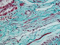
Result
Nuclei |
Blue |
Collagen |
Green |
Muscle |
Red |
Cytoplasm and Erythrocytes |
Red |
Photo x10 Masson Trichrome