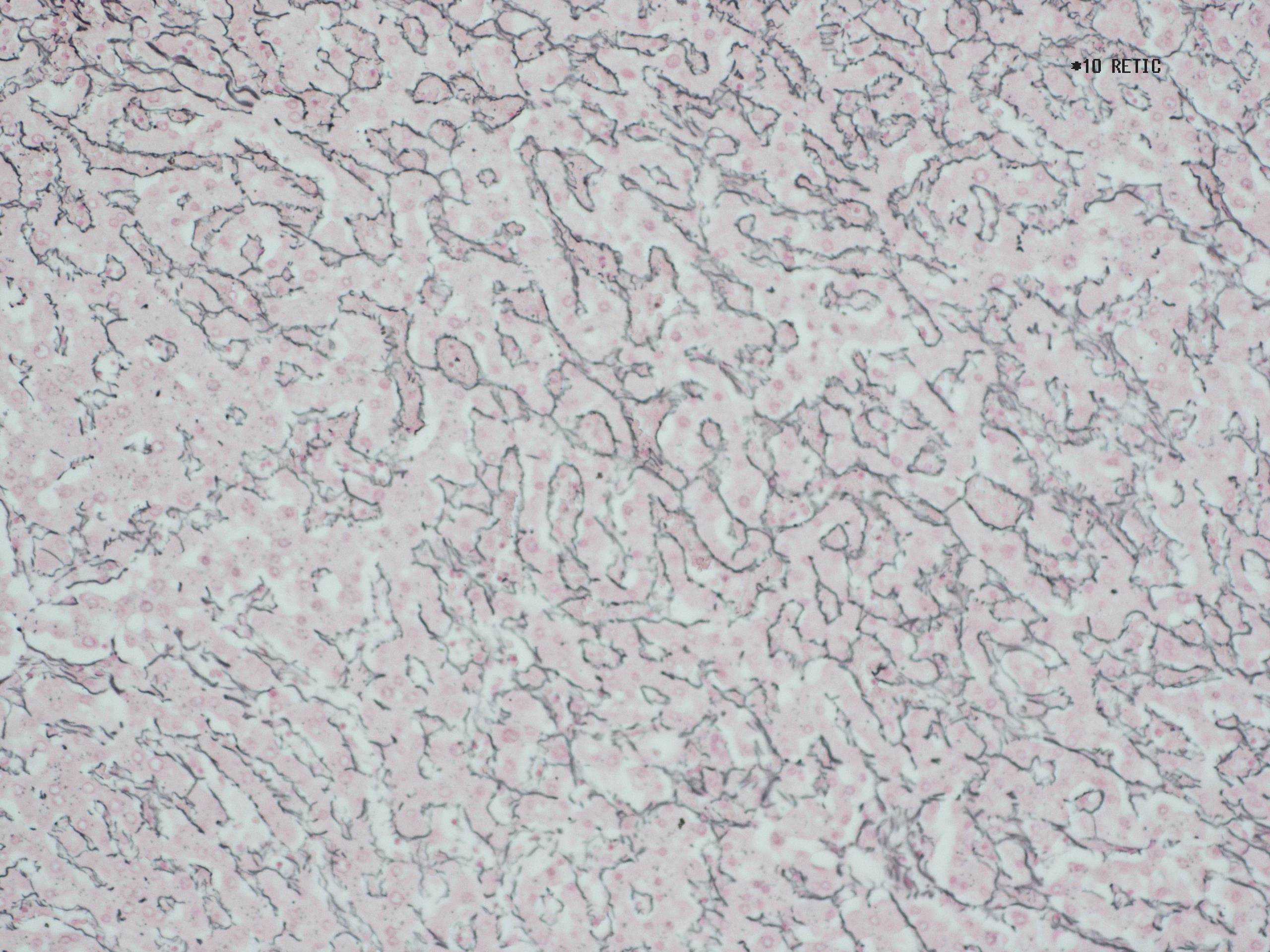
Special Stains - Gordon Sweet Reticulin
- Gordon sweet reticulin background
- Reticulin Technique
Gordon and Sweet's technique demonstrates reticulin fibres using an argyrophil-type reaction involving silver solutions. These techniques rely on the impregnation of the fibres by silver ions and subsequent reduction of these silver ions to their visible metallic form.
A breakdown of the normal reticulin fibre framework can be indicative of several pathological conditions, including cirrhosis and lymphoma. Reticulin stains are often carried out routinely on Liver biopsies as well as bone marrow trephines for this reason.
For this method, there are several different techniques used.
Method
- Take sections to water.
- Treat with acidified potassium permanganate working solution for 5 minutes
- Wash in water, bleach in 5% aqueous oxalic acid for 1 minute
- Wash in water. Treat with 2.5% aqueous iron alum for 15 minutes
- Wash well in several changes of distilled water
- Treat with working silver solution for 1 minute
- Wash well in several changes of distilled water
- Reduce in 10% aqueous formalin for 1 minute
- Wash in water. Treat with 0.2% aqueous gold chloride for 5 minutes
- Wash in water. Treat with 5% aqueous hypo for 3 minutes
- Wash in water. Counterstain in 0.1% nuclear fast red in 5% aluminium sulphate for 5 minutes
- Wash in water, dehydrate, clear and mount
Result
Reticulin Fibres |
Black |
Nuclei |
Red |
Photo - x10 Reticulin
